Predicting Outcomes in Pediatric Hemorrhagic Stroke with Personalized Connectomics
Principal Investigator
Pratik Mukherjee, MD, PhD
Director of the UCSF Center for Imaging of Neurodegenerative Disease and Neural Connectivity Laboratory
Research Coordinator
Shivani Mahuvakar
Study Coordinator, Project 2 at the UCSF Center of Excellence in Hemorrhagic Stroke Research
About Project 2
Connectomics is the study of the brain’s structural and functional connections. The brain is organized with functions located in certain areas. When a hemorrhagic stroke, or bleeding, occurs in a particular area of the brain, the associated function may be lost. For example, bleeding into the language area can cause difficulty in speaking. There are some common trends, but the organization for each person is very different. This makes it difficult to predict the effects of a hemorrhagic stroke for an individual, especially when only the general location of the abnormal blood vessels that caused the bleeding is known. New types of imaging can help us to understand the organization of a person's brain. Project 2, Predicting Outcomes in Pediatric Hemorrhagic Stroke with Personalized Connectomics, will use resting-state fMRI to gather information about pediatric patients’ functional networks, or brain organization. The aim of this study is to determine how an individualized understanding of those networks can help predict the effects of hemorrhagic stroke in children and guide surgical planning to improve treatment outcomes and recovery.
In Project 2 we will assess the utility of advanced functional and structural imaging to predict outcomes after hemorrhagic stroke in pediatric patients. Currently our ability to predict recovery after hemorrhagic stroke is limited to interpreting standard anatomic imaging in the context of expected functional localization derived from population level averages of neurotypical individuals. This has significant implications when treating patients on an individual basis, as opposed to population level average of typically developed adults. Therefore, we will take a novel approach known as lesion network mapping to analyze patient specific connectomics to test our ability to predict outcomes in pediatric hemorrhagic stroke.
We will use resting state functional MRI (rs-fMRI) to study the organization of resting state networks (RSNs) as well as diffusion tensor imaging (DTI) to perform tractography studies of white matter connections between brain regions. RSNs will be the basis of a functional connectome and tractography will be the basis of a structural connectome. These imaging modalities have the unique advantage of being easily obtained alongside routine clinical care resulting in absolute minimal burden to the patient. We will then repeat these measures at serial timepoints throughout recovery, beginning with initial presentation and following along standard clinical follow up. The patient specific connectome will provide a metric with which we will correlate neurologic deficits. We anticipate this will provide greater precision and more personalized care beyond the current standard using anatomic studies alone and inferences based on population level averages. We then will evaluate changes in the functional and structural connectome by analyzing trends in the strengths of these connections over time and in correlation with changes in standardized outcomes metrics. The imaging timepoints will be synergistic with the those in Project 1 and the outcomes metrics synergistic with those in Project 3. The strength of personalized connectomes and within subject comparisons over time will provide a tremendous opportunity to improve the quality of care to this unique patient population. We anticipate these data will provide a strong basis to improve our ability to predict recovery, anticipate sustained deficits, and guide specific therapy based on the needs as predicted by personalized functional and structural connectomes.
MRI Scan of Left Occipital Hemorrhage
Figure 1
Atlas of Resting State Networks (RSN)
Figure 2
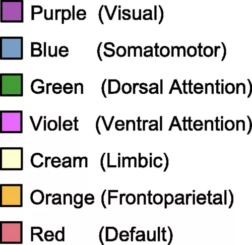
Legend for Atlas of RSN
Figure 2
The hemorrhage demonstrated in Figure 1 is expected to disrupt the Visual RSN as shown in Figure 2.
RSN Atlas Image Source: Yeo BT, Krienen FM, Sepulcre J, Sabuncu MR, Lashkari D, Hollinshead M, Roffman JL, Smoller JW, Zöllei L, Polimeni JR, Fischl B, Liu H, Buckner RL. The organization of the human cerebral cortex estimated by intrinsic functional connectivity. J Neurophysiol. 2011 Sep;106(3):1125-65. doi: 10.1152/jn.00338.2011. Epub 2011 Jun 8. PMID: 21653723; PMCID: PMC3174820.